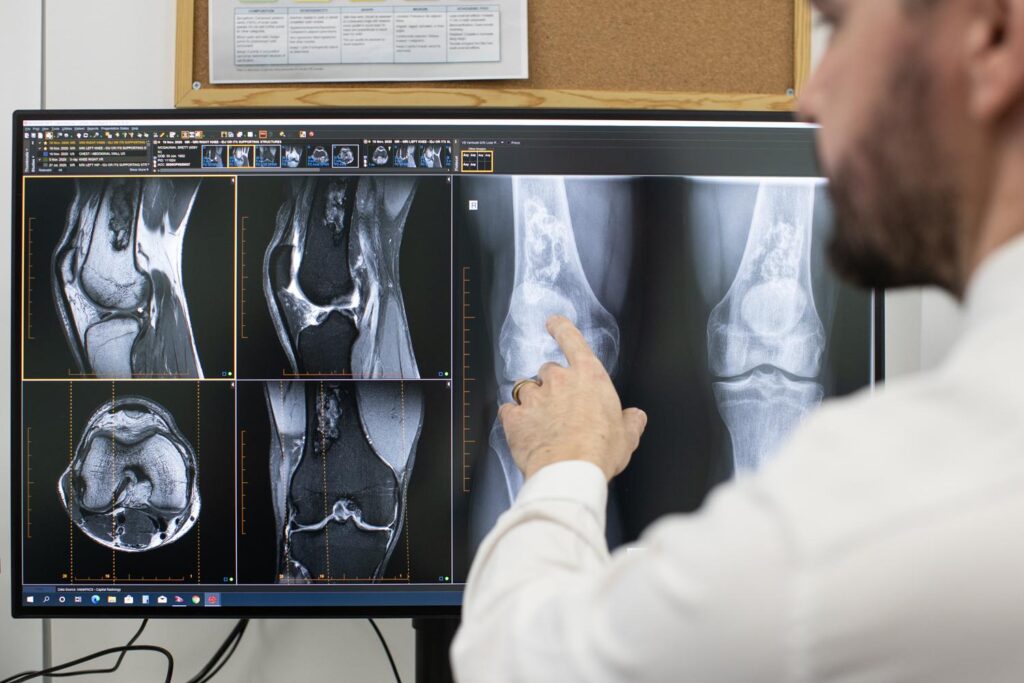
As medical imaging continues going digital, radiologists routinely use software-based tools to view online X-ray reader, MRI scans, and other images.
However, ensuring dimensional measurements onscreen accurately reflect anatomy is critical yet complex.
This guide covers key aspects of digital viewers that radiologists should understand to avoid measurement errors that could impact diagnosis and treatment planning.
Why does Measurement Accuracy Matter?
When interpreting bone scans, one of the most common uses of radiology measurements is assessing fracture healing progress. Surgeons rely on precision within 1-2mm to:
- Decide the best treatment options
- Determine the appropriate timing for surgery
- Evaluate the likelihood of complications
Other examples requiring accurate measurements include:
- Determining malignancy in lesions/tumors
- Sizing implanted devices pre-surgery
- Monitoring growth abnormalities in pediatric cases
Imprecise measurements can directly compromise patient care.
Challenges Measuring Digital X-Ray Images
Traditional analog X-rays provided 1:1 anatomical correspondence where distances on film matched reality. However:
- Digital images involve magnification – The extent depends on how the image was acquired and displayed.
- Pixel displays have finite resolution – Soft tissue contrast differences get lost.
- Window/level settings alter perception – Making tissue appear larger/smaller.
- Software calibrations may be inaccurate – Or not adjusted for display conditions.
Understanding these factors is key for accurate online readings.
Requirements for Precision Image Measurements
Here are four core requirements for achieving precision within 1-2mm when using digital x-ray viewers:
Pixel Size – Resolution should match the capability of the imaging hardware used.
- The minimum pixel pitch should be <200 micrometers.
- Bit depth impacts measurable contrast differences.
High-Fidelity Displays – Monitor size and maximum luminance affect perception.
- The minimum size is 21 inches diagonally.
- Maximum luminance should be >250 cd/m^2.
Calibrated Software – Includes measuring tools reflecting equipment and conditions.
- Measurement tools should calibrate to known magnification factors.
- Calibrate window/level presets to avoid rescaling.
Standardized Protocols – Consistency in acquisition, processing, and display is key.
- Document and follow protocols rigorously.
Below are recommended medical-grade displays for precision diagnosis.
| Model | Luminance | Resolution | Size | Calibration |
| EIZO RadiForce RX1260 | 1450 cd/m^2 | 4MP | 58 cm | DICOM |
| Barco Coronis Fusion 6MP DL | 1000 cd/m^2 | 6MP | 54 cm | Yes |
| Sony LMDX310MD | 500 cd/m^2 | 2MP | 31 cm | Yes |
Investing in proper reading room conditions gives radiologists the tools needed for precise image measurements.

Improving Measurement Consistency
Beyond display and software factors, consistency in how measurements are made also improves accuracy:
- Standard anatomical reference points – Use identifiable bone landmarks.
- Perpendicular angle measurements – Match angle of acquisition.
- Set scale at image level – Avoid relying on client zoom.
- Detailed documentation – Note scales, angles, and landmarks used.
Finally, radiologists should compare prior examinations whenever possible, reviewing changes in anatomical structures over time rather than relying on individual assessments.
The software also increasingly provides advanced measurement approaches like multiplanar reconstructions from CT and MRI volumetric datasets. However, following protocols consistently remains essential.
Action Plan for Accuracy
For medical practices using digital X-ray viewers, ensure accurate, reliable measurements by:
- Reviewing requirements for precision diagnosis
- Comparing display options to find high-fidelity models
- Assessing viewing software measuring tools, calibrations, and consistency
- Establishing and documenting measurement, display,y and reading protocols
- Training all staff on proper precision diagnosis techniques
Getting metrics right is crucial for the correct interpretation of digital images. By understanding what delivers meaningful precision and translating that into robust solutions and workflows, radiologists can feel confident readings reflect reality. Patient health depends on it.


